
Microscopic Root Canal Treatment End Tooth Pain in One Session

Tooth pain can take over your whole day. Eating becomes difficult. Sleep gets interrupted. Even a sip of cold water can send a sharp signal through your jaw. For many people, the words “root canal” add another layer of stress because they imagine pain, long visits, and repeated appointments.
Modern dentistry has changed that experience.
With microscopic root canal treatment, the dentist can see the inside of the tooth in far greater detail, clean the infected or inflamed nerve tissue with more control, and seal the canals with better accuracy. In many suitable cases, this makes it possible to remove the source of pain in one focused visit.
This article explains what the procedure is, how the microscope helps, what happens during the appointment, and why it can be a better choice for saving a painful natural tooth.
Why tooth pain often means the nerve needs treatment
A tooth is not solid all the way through. Beneath the enamel and dentin, there is a soft inner tissue called the pulp. This area contains nerves, blood vessels, and connective tissue. When the pulp becomes inflamed or infected, pain can become intense.
Common causes include:
Deep decay that reaches the nerve
A cracked or fractured tooth
Repeated dental work on the same tooth
Trauma from an injury or strong bite
A deep old filling that no longer protects the tooth well
When the nerve is irritated, the pain may come and go at first. It can later become constant, throbbing, or hard to locate. Some people feel pain when chewing. Others notice sensitivity to hot or cold that lingers after the trigger is gone.
If the infection spreads beyond the tooth root, swelling or a gum abscess may appear. At that point, pain relief alone is not enough. The source of the infection needs to be treated.
A root canal does that by removing the damaged pulp, cleaning the inner canals, disinfecting the space, and sealing it. The goal is simple: save the natural tooth and stop the pain at its source.
What microscopic root canal treatment means
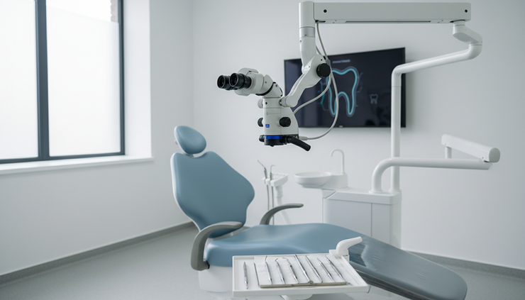
A Microscopic Root Canal uses a dental operating microscope during diagnosis and treatment. This microscope gives strong magnification and bright lighting, allowing the dentist to see details inside the tooth that are too small for the naked eye.
Root canals can be extremely narrow. Some teeth have curved canals, hidden canals, extra canals, or calcified areas where the canal has become harder to detect. Without magnification, these details can be difficult to manage.
The microscope helps the dentist see:
Tiny canal openings
Cracks in the tooth structure
Old filling material inside the canal
Narrow or blocked canals
Curved canal pathways
Areas that still need cleaning
The quality of the final seal
This level of visibility can make treatment more accurate and more conservative. The dentist does not need to remove extra tooth structure just to “search” for the canal. Instead, the microscope guides the work.
That matters because preserving natural tooth structure helps the tooth remain stronger after treatment.

How the microscope helps end pain in one session
The main reason root canal treatment relieves pain is that it removes the inflamed or infected tissue inside the tooth. The microscope improves that process by helping the dentist work with better vision from the start.
For suitable cases, this can shorten the treatment path and reduce the need for multiple visits.
The problem is found faster
The first step in treatment is identifying which tooth is causing pain and what is happening inside it. A dental exam and imaging help guide the diagnosis, but the microscope adds another level of detail during the procedure.
With magnification, the dentist can locate canal openings more quickly and confirm the shape of the chamber inside the tooth. This is especially helpful in molars, which often have multiple canals.
A missed canal can allow infection to remain. Better visibility lowers that risk because the dentist is less likely to overlook small canal openings.
The canals are cleaned more completely
Once the tooth is numb and isolated, the dentist removes the damaged pulp tissue. Then the canals are cleaned, shaped, and disinfected.
The microscope helps during this step because it gives the dentist a clear view of the canal entrance and internal tooth structure. Fine instruments can then be used with more control.
A cleaner canal system means fewer irritants remain inside the tooth. That is a key reason pain can settle after treatment.
The seal is placed with greater accuracy
After cleaning, the canals are filled and sealed with a biocompatible material. The seal helps stop bacteria from re-entering the tooth.
A precise seal is one of the most important parts of long-term success. If gaps remain, bacteria can return and cause future pain or infection. The microscope helps the dentist check the area carefully before completing the filling.
In many cases, this careful sequence allows the painful tooth to be treated in a single appointment. Some cases still need more than one visit, especially when there is severe infection, swelling, complex anatomy, or retreatment of a previous root canal. A proper exam is the only way to know.
The purpose of microscopic treatment is not to rush the appointment. It is to do the work more accurately, so the source of pain can be treated as completely as possible.
What happens during a one-session appointment
A root canal visit feels less mysterious when the steps are clear. While every case is slightly different, most microscopic root canal appointments follow a structured process.
The dentist confirms the diagnosis
Before treatment begins, the dentist checks the tooth, reviews symptoms, and may take dental imaging. This helps confirm that root canal treatment is the right choice and that the tooth can be saved.
Not every toothache needs a root canal. Gum problems, sinus pressure, bite issues, or another nearby tooth can sometimes cause similar symptoms. A careful diagnosis prevents the wrong treatment.
The tooth is fully numbed
Local anesthesia is used so the area becomes numb before the procedure starts. The goal is comfort during treatment. Many patients are surprised that the appointment feels similar to having a filling, only longer and more detailed.
If anxiety is high, talking with the dental team before the appointment can help. They can explain how breaks, communication signals, and comfort options work.
The tooth is isolated and protected
The dentist usually places a protective sheet called a dental dam around the tooth. This keeps the area dry and reduces saliva contamination during treatment.
A clean, dry field helps the dentist disinfect and seal the canals properly.
The microscope guides the cleaning
The dentist creates a small opening in the tooth to reach the pulp chamber. The microscope is then used to locate the canal openings and guide the cleaning process.
Fine instruments clean and shape the canals. Disinfecting solutions help flush out bacteria and tissue remnants.
Because the microscope improves vision, the dentist can work with more confidence in narrow or complex areas.
The canals are filled and sealed
After cleaning and shaping, the dentist fills the canals and seals the space. This step helps protect the tooth from reinfection.
Depending on the tooth, a temporary or permanent filling may be placed at the end of the appointment. Many back teeth later need a crown because they handle strong chewing forces. The dentist will explain the restoration plan based on how much tooth structure remains.

Why choose microscopic treatment for a painful tooth
The main benefit is accuracy. When the dentist can see more, they can treat more precisely. That can affect comfort, cleaning quality, and long-term tooth preservation.
Here is how microscope-assisted treatment compares with a traditional approach in practical terms:
Better visibility
The microscope enlarges the treatment area and improves lighting inside the tooth.
Less unnecessary tooth removal
The dentist can identify canals without widening the access more than needed.
Helpful in complex cases
Calcified canals, retreatments, and unusual canal shapes can be easier to handle.
More precise cleaning
Hidden canals, narrow spaces, and curved areas can be managed with greater control.
Improved sealing control
The final filling can be checked more carefully before the tooth is restored.
Fewer avoidable complications
Clearer vision may reduce the risk of missed canals or incomplete cleaning.
For patients in Cairo and across Egypt looking for advanced endodontic care, this approach can be especially valuable when pain is severe and time matters. A well-planned one-session treatment can reduce repeated clinic visits and help patients return to eating, sleeping, and daily life with less disruption.
Still, one-session care must be chosen case by case. The dentist will look at infection level, swelling, tooth anatomy, previous dental work, and the final restoration needed. If a second visit gives a better result, the dentist should recommend it.
The best treatment is not the fastest one. It is the one that removes the source of pain and gives the tooth the best chance to last.
What to expect after treatment
Some tenderness after a root canal is normal, especially when biting. The tooth and surrounding ligament may need time to calm down after being cleaned and sealed. This discomfort is usually manageable and should improve as healing begins.
The dental team may recommend pain relief instructions based on your health history. Follow only the medication plan given by your dentist or physician.
After treatment, protect the tooth by avoiding hard chewing on that side until the final restoration is complete. If the tooth needs a crown, do not delay it. A root canal-treated tooth can still crack if it is not properly protected.
Call the clinic if pain gets worse instead of better, swelling appears, the temporary filling comes out, or the bite feels too high. These issues can often be corrected quickly when addressed early.

FAQ
Is microscopic root canal treatment painful?
The tooth is numbed before treatment, so the procedure should not feel painful. Some pressure or vibration may be felt. Mild soreness after the visit can happen, but it usually improves as the tooth heals.
Can every root canal be finished in one session?
No. Many cases can be completed in one session, especially when the tooth is suitable and infection is controlled. Severe swelling, complex anatomy, or retreatment may require more than one visit.
Why is a microscope better than regular vision?
The microscope magnifies the inside of the tooth and adds strong lighting. This helps the dentist find small canals, clean narrow spaces, and check the final seal more accurately.
Will the treated tooth need a crown?
Many back teeth need a crown after root canal treatment because they handle heavy chewing forces. Front teeth may not always need one. The decision depends on tooth strength, decay size, and how much natural structure remains.
How long can a tooth last after treatment?
A well-treated and properly restored tooth can last for many years. Good oral hygiene, regular checkups, and a strong final restoration all help protect the result.
Take control of tooth pain before it controls your day
Root canal treatment no longer has to mean fear, repeated visits, or drawn-out discomfort. With microscope-assisted care, the dentist can see tiny details, clean the canals with greater accuracy, and seal the tooth carefully.
For the right case, that can mean relief from severe tooth pain in one session while preserving the natural tooth.
If tooth pain is affecting your sleep, meals, or daily routine, do not wait for it to get worse. Book your consultation with ADC Dental Clinics and find out whether microscopic root canal treatment is the right solution for your tooth.
This article is for general educational purposes and does not replace a dental exam or personalized medical advice.


Comments