
Microscopic Dentistry Guide for Precise and Painless Root Canal Care

A root canal has a reputation it no longer deserves. For many people, the phrase brings up thoughts of pain, long appointments, and uncertainty about whether the tooth can really be saved. Modern root canal care is very different, especially when a dental microscope is part of the treatment.
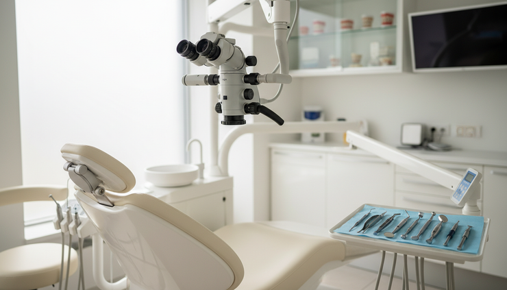
Microscopes give dentists a magnified, well-lit view of the inside of the tooth. That matters because root canals are tiny, curved, and often more complex than they appear on an X-ray. When the dentist can see more clearly, they can clean more precisely, preserve more healthy tooth structure, and treat problems that may be missed with the naked eye.
This guide explains how Microscopic Dentistry supports more precise and comfortable root canal treatment, why it can improve the chance of saving a natural tooth, and what to expect during care at a modern dental clinic such as ADC Clinics in Cairo.
This article is for general dental education only. A dentist or endodontic specialist should examine the tooth before recommending any treatment.
Why root canals need such a close look
A tooth is not solid all the way through. Inside it is a soft tissue space called the pulp, which contains nerves and blood vessels. When deep decay, cracks, trauma, or repeated dental work irritates or infects the pulp, a root canal may be needed.
During a root canal, the dentist removes infected or inflamed tissue, cleans the canal system, shapes the space, and seals it. The goal is to stop infection while keeping the natural tooth in place.
The challenge is scale. Root canals can be extremely narrow. Some teeth have extra canals, branching areas, old filling material, calcified sections, or curved roots. A molar, for example, may have several canals that do not run in straight lines.
Without magnification, the dentist relies on experience, X-rays, touch, and standard lighting. Those tools are still important, but they do not always show the fine details inside the tooth. A microscope adds a major advantage: direct visual precision.
Better visibility helps the dentist identify:
Small canal openings
Hairline cracks in the tooth
Hidden decay under old restorations
Blocked or calcified canals
Remaining infected tissue
Previous root canal complications
Tiny perforations or structural defects
This is why microscopes have become an important part of advanced endodontic care. They help the clinician treat what is actually there, not just what is expected to be there.
How microscopic dentistry improves root canal precision
A dental operating microscope does two main things. It magnifies the tooth, and it focuses bright light directly into the working area. That combination changes the way a dentist approaches root canal treatment.
It helps locate hidden canals
Missing a canal is one of the reasons a root canal can fail or require retreatment. Some canals are very small, partially covered by calcified dentin, or positioned in unexpected areas.
Under magnification, the dentist can study the floor of the tooth chamber more carefully. Subtle color changes, grooves, and anatomical patterns become easier to see. This helps guide the search for canals without unnecessary drilling.
That matters because finding the canal is only one part of the goal. The dentist also wants to preserve as much natural tooth structure as possible.
It allows more conservative treatment
Precision is not just about doing more. Often, it is about doing less damage.
When the dentist can see exactly where to work, they can make smaller adjustments and avoid removing healthy tooth structure. This is especially important in teeth that are already weakened by decay, cracks, large fillings, or previous treatment.
A more conservative approach may support the long-term strength of the tooth. In many cases, the tooth will still need a crown or final restoration after root canal therapy, but preserving structure gives that restoration a better foundation.
It supports cleaner canal preparation
A root canal succeeds when the infected space is thoroughly cleaned, shaped, disinfected, and sealed. Microscopes help the dentist inspect the access area and canal openings throughout the procedure.
They also help during complex steps such as removing old filling material, managing a separated instrument, or checking for tissue remnants in the chamber. While no tool can guarantee a result, improved visibility can reduce uncertainty and support a cleaner, more controlled treatment.

Why treatment can feel more comfortable
Many people connect root canals with pain, but root canal treatment is designed to relieve pain caused by infection or inflammation. The procedure itself is usually done with local anesthesia, so the tooth should be numb during treatment.
Microscope-assisted care may also improve comfort in indirect ways.
When the dentist can see clearly, they can work with more confidence and control. That can reduce unnecessary probing, repeated searching, or excessive removal of tooth structure. The appointment may still take time, especially for complex teeth, but the process tends to be more focused.
A microscope also helps the dentist detect cracks or complications earlier. If a tooth has a vertical root fracture, for example, saving it may not be possible. Identifying that sooner can prevent repeated procedures that would not solve the problem.
Comfort also comes from communication. A good clinical team explains what they see, why treatment is needed, and what the patient may feel afterward. Mild soreness after a root canal can happen, especially when the tooth was infected before treatment. It usually improves as the area heals, and the dentist may recommend medication or follow-up care based on the situation.
The key point is simple: pain control starts with diagnosis, anesthesia, and careful technique. Microscopic magnification supports all three by helping the dentist understand the tooth in greater detail.
How microscopes help save natural teeth
A natural tooth is worth saving when it can be restored predictably and safely. Root canal treatment exists for that reason. It gives a damaged or infected tooth a chance to remain in function instead of being removed.
Microscopes can improve that chance in several ways.
They make retreatment more possible
Some patients come in with a tooth that had a root canal years earlier but now shows pain, swelling, or infection. Retreatment may be needed if bacteria remained inside the canal system or entered again through a leaking filling or crown.
Retreatment can be complex. The dentist may need to remove old material, identify missed canals, clean around posts, or manage scarred and narrowed pathways. Magnification helps guide these steps with more accuracy.
This does not mean every failed root canal can be saved. Some teeth are too cracked, too loose, or too damaged. Still, microscopic treatment can make more informed decisions possible.
They help manage difficult anatomy
No two teeth are exactly alike. A textbook diagram may show three canals, but the tooth in the chair may have four. A canal may split, curve sharply, close off with calcification, or hide beneath a ledge of hard tissue.
Microscopic Dentistry gives the dentist a better chance of recognizing those variations. That can affect the entire outcome of treatment.
A missed canal can allow infection to continue. A poorly cleaned area can become painful later. A hidden crack can change the treatment plan completely. Seeing small details early helps the dentist choose the right approach before problems grow.
They support better sealing and final restoration planning
Root canal therapy does not end with cleaning. The canals must be sealed, and the tooth must be restored to prevent bacteria from re-entering.
Magnification helps the dentist inspect the internal space before sealing. It can also reveal whether the remaining tooth structure is strong enough for a filling, inlay, onlay, or crown. That connection between root canal treatment and final restoration is important. A well-treated canal still needs a strong seal above it.

What to expect during a microscope-assisted root canal
A microscope-assisted root canal feels similar to a conventional root canal from the patient’s point of view. The main difference is the level of detail the dentist can see while working.
A typical visit may include the following steps.
Clinical exam and imaging
The dentist checks the tooth, reviews symptoms, and takes X-rays or other imaging if needed. The goal is to confirm whether the pulp is inflamed or infected and whether the tooth can be restored.
Local anesthesia
The tooth and surrounding area are numbed before treatment starts. Patients who feel anxious should tell the dental team before the appointment. The team can explain comfort options and pause when needed.
Isolation with a dental dam
A small protective sheet called a dental dam is often placed around the tooth. It helps keep the tooth dry and reduces saliva contamination during the procedure.
Microscope-guided access
The dentist creates a small opening to reach the pulp chamber. Under magnification, they locate canal openings and inspect the internal tooth structure.
Cleaning, shaping, and disinfection
Special instruments clean and shape the canals. The dentist uses disinfecting solutions to reduce bacteria inside the canal system.
Sealing the canals
After cleaning, the canals are filled and sealed with dental material. In some cases, treatment may take more than one visit, especially if infection is active or the tooth is complex.
Final restoration
A permanent filling or crown protects the tooth after treatment. Back teeth often need crowns because they handle heavy chewing forces.
Patients should follow aftercare instructions closely. Avoid chewing hard foods on the treated tooth until the final restoration is complete. Call the clinic if pain worsens, swelling develops, or the bite feels too high.
When microscopic root canal care is especially helpful
Microscope-assisted dentistry can help in many root canal cases, but it is especially valuable when the tooth is complicated.
It may be recommended for:
Molars with several canals
Teeth with narrow or calcified canals
Retreatment of an old root canal
Suspected missed canals
Cracked tooth evaluation
Removal of old posts or materials
Cases where previous treatment did not heal as expected
Teeth with unusual root shape or canal anatomy
It can also help when the goal is to preserve a tooth that might otherwise seem difficult to save. Clear vision helps the dentist judge whether saving the tooth is realistic or whether another option, such as extraction and replacement, would be more appropriate.
For patients in Cairo, access to microscope-assisted care at ADC Clinics means complex root canal treatment can be planned with modern diagnostic thinking and close visual control.
FAQ
Is a microscopic root canal painful?
The procedure is usually performed with local anesthesia, so the tooth should be numb during treatment. Some soreness after the visit can happen, especially if the tooth was infected, but this is usually manageable with the dentist’s guidance.
Does a microscope make the root canal faster?
Not always. Some cases still take time because the tooth anatomy is complex. The main benefit is not speed. It is better visibility, which supports more careful cleaning and decision-making.
Can every tooth be saved with microscopic dentistry?
No. A microscope can help the dentist see more and treat with greater precision, but some teeth are too fractured, loose, or damaged to restore safely. The exam determines whether saving the tooth is the right choice.
Is microscopic treatment only for retreatment cases?
No. It can help with first-time root canals as well. It is especially useful for molars, narrow canals, calcified canals, and teeth with unusual anatomy.
Will I still need a crown after a root canal?
Many back teeth need a crown after root canal treatment because they are exposed to strong chewing pressure. The dentist will recommend the final restoration based on how much tooth structure remains.

A clearer way to treat and protect natural teeth
Microscope-assisted root canal treatment gives dentistry something powerful: a clearer view of a very small space. That clarity can help the dentist find hidden canals, clean more thoroughly, preserve healthy structure, and make better decisions about whether a tooth can be saved.
For patients, the benefit is practical. Treatment can feel more controlled, more precise, and less intimidating. A root canal is no longer something to fear. With the right technology and the right clinical team, it can be a careful step toward keeping a natural tooth healthy and functional.
If you have tooth pain, swelling, sensitivity, or have been told you may need a root canal, the next step is an exam. Book a consultation with ADC Clinics to discuss whether microscope-assisted root canal care is right for your tooth.


Comments